

Having dental implant treatment is often more complex than people may initially think.
Let’s take a look at dental implants in the front of the mouth. Often teeth in this area are lost because of trauma, or because they have had all sorts of treatments already carried out (root treatments, posts, crowns etc), and now we are out of options and need to look at the removal of the tooth (or teeth)
This blog post does not apply to teeth that have been lost to gum disease – This is a different problem.
But let’s have a look at this case, where this patient lost her front teeth many years ago, and she had a bridge to replace them.


It should be noted that a bridge is a great option, and it served her really well for many years
But what we can see is that there is a gap between the bridge and the gum.
This was not there in the beginning, originally, the teeth would have touched the gums.
But when teeth are removed, the bone around the teeth is also often lost over time.
The vast majority of this bone loss happens in the first 3-4 months, but then it continues for years, at a much slower rate.
Having a fixed reference, like the bridge here, really highlights how much bone is lost.
One factor that we need to think about it: is this visually a problem?
If the junction between the gum and the teeth is not visible when smiling or talking, then it is sometimes it is possible to place a dental implant in this position and accept that the look of the tooth will not be perfect if the lips are lifted out of position.
But in this case, we did not have that option, the patient shows a lot of gum when smiling, and this gap was her biggest problem – she was very self-conscious about it.
We could have made a new bridge, but the supporting teeth were not great, so even though she was absolutely terrified, she decided that bone grafting and dental implants were the right long term decision for her.
Let us have a look at the CBCT Scan. This type of scan lets us plan dental implant treatment properly, and it also lets you see how much bone we actually have to play with.
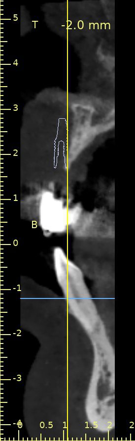
So this image taken from the scan shows us the thickness of the top bone, and there is a simulated dental implant in its ideal position.
The dental implant is completely out of the bone – there is no way to have an implant here without increasing the amount of bone available.
In dental terms, this is a really big volume of bone that we need.
Ideally, we look to have a minimum of 1mm of bone all the way around the implant, this is a 3.5mm implant (width), so really we are looking to have 4-5mm of increased bone. This is a lot in dental terms.
There are a number of ways that we can increase the bone, and I will likely have a post dedicated to this later, but in this case, we mainly took all of the bone from another area of her jaw.
As she was terrified about having any dental treatment, let alone anything this invasive, she opted to have sedation where she was in a kind of twilight zone for the treatment, it lets us do what we needed to, and she didn’t remember anything after the treatment.
Now, this is invasive, there is a lot of swelling and bruising to be expected, in fact, she is documenting her experience on her Instagram account.
Depending on the type of bone graft, it can take between 4-9 months to form good enough bone to place the dental implants.
The advantage of using as much “own bone” is that the healing is better and quicker, the disadvantage is that it’s a more complex procedure and there are two areas that need to heal.
Once the dental implants are in, creating the smile is going to be a lot easier – often these treatments are very intense in the first phases, and then things get easier.
During the healing phase, we are using two types of temporary teeth, one that can be used when there is a lot of swelling, and then once the swelling is reduced, we will make a temporary fixed bridge.


